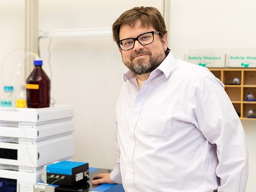
Featured Customer: Darren Begley, Ph.D.

Novel therapeutic modalities are always exciting, and often present new challenges for analytical characterization. That’s where a solid grounding in multiple biophysical techniques comes in handy: understanding the capabilities, strengths and weaknesses of each technique and instrument guides the optimal selection and development of an analytical strategy. Dr. Darren Begley of Beam Therapeutics has seen and analyzed a wide variety of therapeutically-relevant biomolecules with many such techniques. Read on to find out how he utilizes light scattering to characterize gene delivery and other biotherapeutics.
Please tell us about your background: where you grew up, studied and the field you chose.
I grew up in Canada and developed an interest in different kinds of spectroscopy while completing a Joint Honours Chemistry and English Literature program at McGill University in Montreal. I then moved to California, where I first learned to use nuclear magnetic resonance (NMR) spectroscopy to study biomolecular interactions with Maurizio Pellecchia at Triad Therapeutics (now at UC Riverside). I continued that work with my Ph.D. advisor Gabriele Varani at the University of Washington in Seattle, looking at small molecule interactions with proteins and structural (non-coding) ribonucleic acid (RNA) molecules. After spending some time in the structural biology field, I transitioned into analytical development to biophysically characterize a wide array of medically relevant biomolecules. I have investigated short RNA, DNA and synthetic oligos with various linkers, modifications and non-nucleic acid attachments, trying to assess identity, purity and other properties. I have characterized mature mRNA molecules, stable proteins including antibodies, virus particles and other molecular species to determine their overall mass, size, and level of aggregation in solution. I have also analyzed complexes containing 1 or more types of molecules, such as nucleic acid-protein complexes, and investigated lipid nanoparticles and other kinds of drug delivery vehicles. The chief focus of my work has been on pharmaceutically relevant materials, with the goal of helping patients today and tomorrow live better lives.
What does your current position entail? How does it tie into your previous experience, and where is it going?
I work at Beam Therapeutics, a gene therapy company developing a platform to create cures for genetic diseases. We are leveraging CRISPR and nucleobase modification technology from David Liu's lab at Harvard University to manifest what we hope are permanent corrections to disease states in the human genome. These are complicated biological machines which require novel, intricate modes of delivery.
In my career as an analytical chemist, I have focused on biophysical, solution-state methods to characterize biological molecules. These include dynamic light scattering (DLS), multi-angle light scattering (MALS), circular dichroism (CD), analytical ultracentrifugation (AUC), capillary electrophoresis (CE), UV/VIS absorbance, as well as NMR, cryoTEM and mass spectrometry (MS) as part of a larger team. I have found that using multiple orthogonal approaches to characterize something is the way to go. You have much greater confidence in the data when several methods corroborate each other. Conversely, when results from multiple experiments do not add up, you have to ask yourself what is going on, what is causing these differences. Biophysics tools can provide insights which you are not necessarily looking for, but which end up being pretty critical in the grand scheme of things. I also benefit from a generally high level of robustness in these methods; solution-state biophysical tools tend to work most of the time, without much tinkering once they are set up properly. The main challenge is in correctly interpreting the data.
In what context did you first learn about light scattering and Wyatt instruments?
I first started using light scattering to characterize small molecules and proteins for NMR and X-ray crystallography screening campaigns. With small molecules, you cannot always tell which ones are well behaved in solution by just visual inspection. A quick DLS scan will reveal the presence of small molecules which form micelles and/or other large species in solution which are otherwise invisible, something critical to know when studying binding interactions and other fundamental properties.
Knowing the polymeric state of your protein or nucleic acid molecule is also vital information, a characteristic which you cannot be certain of from retention time alone. I have worked on several projects now where the quaternary structure of a given protein (is it a monomer, a dimer, a trimer, etc.) estimated by size-exclusion chromatography (SEC) retention time correlated to a set of external protein standards was flat-out wrong. The protein we thought was (in one case) a monomer was actually a dimer, but because of its compact structure it eluted later than expected. In every single one of these cases, SEC-MALS showed the correct polymeric state of the protein when regular SEC did not. That is a powerful tool, and for me an essential one when probing fundamental molecular characteristics.
How has your Wyatt instrumentation contributed to your research and development studies?
I routinely use DLS and MALS as a "first pass," and continue to advocate for their application in any new project. I really want to know the mass and polymeric state of all preliminary test materials before we spend a lot of time and resources in further studies. It also affords a relatively quick way to inspect samples for potential aggregates or degradants in solution. We cannot see everything by DLS and MALS, but the data provide a great starting point and will often dictate what analytics come next.
I think where MALS with SEC or a field-flow fractionation (FFF) device has a lot to offer the gene therapy community is in characterization of virus particles, a popular vehicle for drug delivery. I am not a virus biologist by training, but I have used light scattering techniques extensively to characterize adeno-associated virus (AAV), as well as other viruses and virus-like particles. When I started working with AAV for gene therapy I was surprised to learn that the field still relies heavily on methods for ratio of empty-to-full particles which are tedious, low-throughput, and not as accurate as desired. But figuring those things out, it is not a trivial task. Empty and full AAV particles tend to be the same size and tend to elute as a single peak by SEC. The AAV shell protein and nucleic acid cargo both absorb UV light, making it difficult to determine how much of each is present by UV absorbance. There are plenty of flow cytometers around, but none of them can count particles in the nanometer diameter size range of most viruses, so even getting a total particle count is not straight-forward.
Over a year ago I developed a keen interest in number density determinations (i.e. particle concentration) by MALS to address some of these concerns, and started working with Wyatt researchers and application support staff to see if light scattering could help. Preliminary results have shown accurate masses for samples consisting of mainly empty virus particles vs full ones, while both retain the same overall size, as determined by MALS with a QELS unit. There are others working on solutions as well, incorporating ion exchange chromatography to get empty-full AAV separation for some serotypes. A recent poster by Paul Gagnon and others at BIA Separations reveal a MALS detector with an appropriate column and salt gradient can provide more accurate empty:full ratio than simple UV, using fluorescence data as reference. So right now I am pretty excited to be working on these new analytical challenges, and I look forward to working with folks at Wyatt on MALS-based approaches to rapidly generate useful characterization data for virus particles and other novel materials for drug delivery here at Beam.

Wyatt’s SEC-MALS and field-flow fractionation instruments have a lot to offer the gene therapy community in characterization of virus particles.
