How can I tell if I have fluorescing or absorbing samples?

Introduction
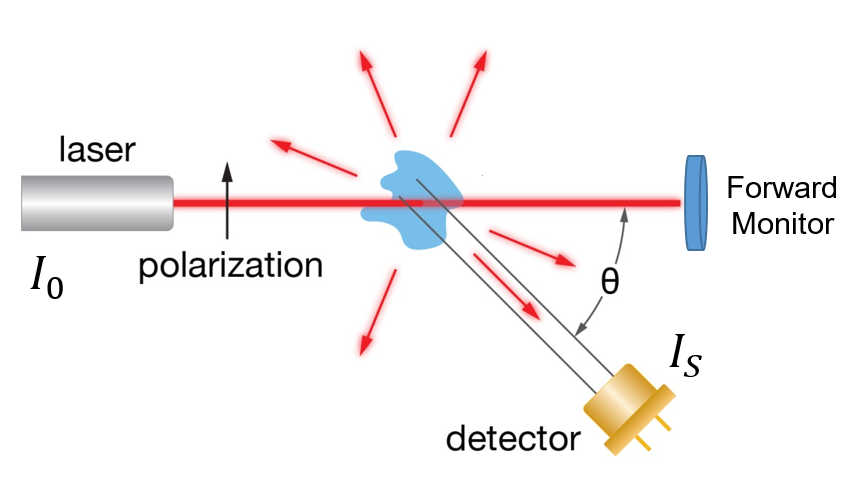
Multi-angle light scattering (MALS) analysis is based on the intensity of scattered light as measured by an array of photodiodes. To accurately calculate the molar mass of a sample, we need to account for any excess light (fluorescence) or a decrease in the intensity of the laser source (absorbance) so these changes aren’t incorrectly attributed to the sample being analyzed.
Signs to look for:
- Absorption of laser light as detected by the instrument forward monitor
- Unexpectedly high molar mass increasing with elution volume
The default MALS laser wavelength for the DAWN®, miniDAWN®, and microDAWN™ is around 658 nm, which for many samples completely avoids the excitation band. If the sample’s absorbance in the red region is unknown, looking at an ASTRA experiment file can determine if absorbance occurs and then correct for it.
Samples that absorb light in the laser wavelength of the MALS detector show an artificially low molecular weight since the software uses the laser monitor signal (I0) to normalize the scattering signals relative to incident laser beam power.
Diagnosing absorption: Basic Collection
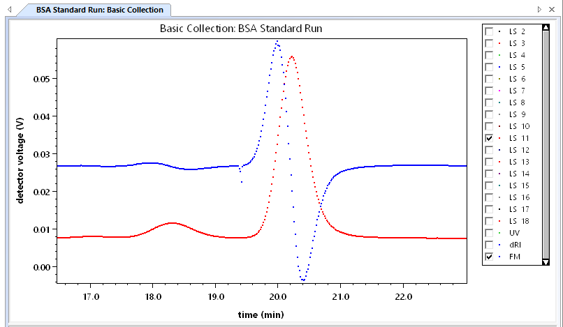
To diagnose if your sample is absorbing the laser light, first open the Basic Collection procedure for the ASTRA experiment file. Enable only the 90° detector (LS 11 for a DAWN, LS 2 for a miniDAWN or microDAWN) and the forward monitor (FM). If the forward monitor signal appears to be an inverted or flipped copy of the light scattering signal, while maintaining alignment of the two signals, it is likely that absorption is occurring.

Diagnosing absorption: Forward monitor
During measurement of non-absorbing samples there will likely be a small effect on the forward monitor signal due to the sample flowing into the flow cell, however, this signal will not mirror the light scattering signal.
The forward monitor voltages are not displayed to scale in the Basic Collection procedure. To view the voltages of the FM signal, hold down the Shift key while pressing the left mouse button. Sample absorbance may be corrected if the forward monitor does not drop by more than approximately 20%.
Correcting for Absorption
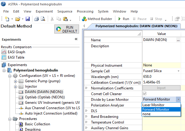
To correct for a sample which absorbs the laser light, open the ASTRA experiment file and open the Configuration for your MALS detector. Select Forward Monitor from the Divide by Laser Monitor drop down menu and click OK.
Note: Selecting to divide by the “rear” laser monitor is the preferred and recommended option. The forward monitor should only be selected when the sample has been determined to absorb light at the operating wavelength of the laser.
Conclusion
The forward monitor voltages are not displayed to scale in the Basic Collection procedure. To view the voltages of the FM signal, hold down the Shift key while pressing the left mouse button. Sample absorbance may be corrected if the forward monitor does not drop by more than approximately 20%.
Some absorbing samples, such as polymerized hemoglobulin in this example, absorb the laser light but don’t fluoresce. In other samples, correcting for absorbance may not be enough if they exhibit fluorescence during the experiment. So how can you tell if your sample is fluorescing? If a sample is fluorescing, it will show a suspiciously high molar mass increasing with elution volume as shown below.
We’ll discuss strategies for using light scattering techniques with fluorescing samples in our next Ask the Expert. Do you have a question? Contact our experts here in Customer Support. We’re happy to help! Call +1 (805) 681-9009 option 4.
